Thoracic Stenosis | Myelopathy Specialist
Spinal cord compression can happen to anyone. Over time, wear and tear, aging, gravity, the thickening of ligaments, or a traumatic injury, can all lead to this condition. Although not life-threatening, it is a serious condition that can lead to permanent disability or paralysis. Conservative measures can help relieve symptoms, but sometimes a more comprehensive treatment may be required. If you are experiencing symptoms of spinal cord compression, it is recommended you see a specialist immediately. Doctor Andre M. Samuel, orthopedic spinal doctor, treats patients in Clear Lake, Houston, Sugar Land, TX area who have been diagnosed with thoracic stenosis or myelopathy. Contact Dr. Samuel’s team today.

What is thoracic stenosis?
The spine is composed of 33 bones stacked on top of each other that run from the cranial base (bottom of the skull) to the tailbone. These bones are known as vertebrae. But the spine is composed of more than just these vertebrae. Running down the center of each vertebra is a hollow space that houses the spinal cord. This space, known as the spinal canal, also contains 31 pairs of nerve roots communicating motor, sensory, and other impulses with the rest of the body.
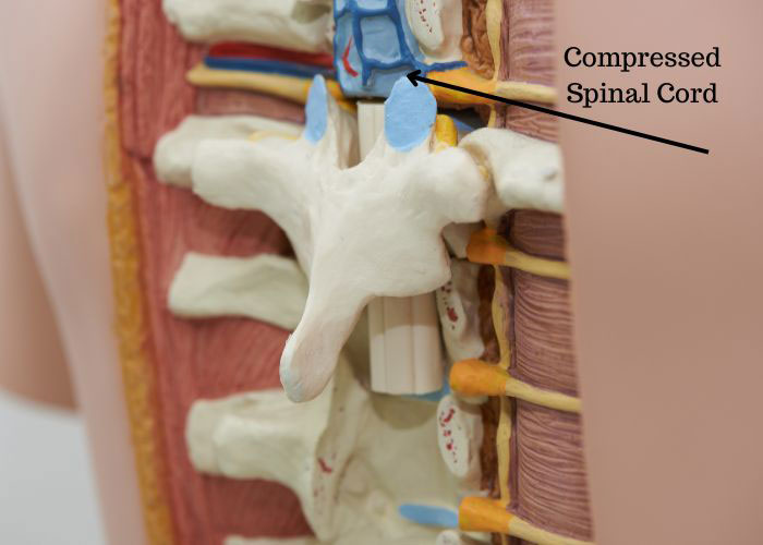
The areas of the back are generally considered in four major areas: the cervical (neck), the thoracic (mid-back), the lumbar (low-back), and the sacral/coccygeal (tailbone) regions. Sometimes, the space in the spinal canal can be reduced in volume, causing a narrowing of the spine known as stenosis. When the spinal canal is compressed in the thoracic region, it can pressure the spinal cord or the nerves in the area that radiate a sharp pain to other parts of the body. This is called thoracic stenosis. Stenosis of the spine can result in a neurological condition known as myelopathy.
Thoracic stenosis (also known as spinal cord compression or thoracic myelopathy) is a serious condition that can cause poor walking balance, problems with coordination, and bowel dysfunction. Doctor Andre M. Samuel, orthopedic spinal Doctor, treats patients in Clear Lake, Houston, Sugar Land, TX area who have symptoms associated with myelopathy or thoracic stenosis.

What causes thoracic stenosis?
Stenosis can happen to anyone and can occur in any region in the back. Although acute, traumatic events such as a sports injury, disc herniation, or a car accident can lead to this condition; thoracic stenosis is more often caused by the aging process. As we get older, the joints of our spine deteriorate, and the space between our vertebrae shrinks. Other degenerative factors, such as osteoarthritis and bone spurs, can also lead to this condition.
Thoracic stenosis can also be caused by congenital factors. People born with a narrower spinal canal, for example, or those with excessive bone growth, can be more susceptible to thoracic myelopathy.
What are the symptoms of thoracic stenosis or myelopathy?
- Difficulty walking or poor walking balance or coordination
- Pain in the back or legs
- Incontinence or loss of bowel control
- Rib area or internal organ pain
- Numbness, tingling, or weakness in torso or legs
How is myelopathy or thoracic stenosis diagnosed?
To accurately diagnose thoracic myelopathy, Dr. Samuel will conduct a review of your medical history and perform a physical exam. to determine sources of your pain. Imaging tests may also help diagnose the problem. Some common imaging tests may include:
- X-rays(radiographs) of the spine can help determine any arthritis or instability in the spine. It primarily shows us the bony structures of the spine including fractures or injuries. It can also be used to measure spinal alignment/posture.
- MRI’s (magnetic resonance imaging) is advanced technique that does not require radiation to the body that is used to determine any narrowing of the spinal canal and compression of the spinal cord or nerve roots. This can also be used to determine if there is any permanent injury to the spinal cord from spinal cord compression.
- At CT scan (computed tomography) is a combination of multiple X-rays (radiographs) that together recreate a high resolution 3D image of a patients bony anatomy. This is used in some cases to better evaluate a patients bony anatomy and determine whether rare conditions exisit, including OPLL (ossification of posterior longitudinal ligament), dysplastic pedicles (condition making spinal fusion procedures more difficult/dangerous), or congenital anomalies.
- In cases when MRIs cannot be performed (in patients with certain implanted pacemaker or mechanical devices or patients with severe claustrophobia) and special text called a CT Myelogram was be used to measure the amount of spinal canal narrowing and spinal cord compression in suspected cases of thoracic stenosis/myelopathy.
What is the treatment for thoracic stenosis?
A variety of factors help determine the best course of treatment for patients diagnosed with thoracic stenosis or thoracic myelopathy. These treatments can be non-surgical or surgical.
Non-surgical treatment:
The aim of non-surgical treatments for patients is to relieve pain through the reduction of nerve inflammation and pressure. To achieve this, a specialist may recommend:
- NSAID’s, or non-steroidal anti-inflammatory drugs, to reduce pain or inflammation.
- Physical therapy to improve posture and strengthen the muscles of the thoracic area.
- Weight control techniques.
- Epidural spinal injections.
Surgical treatment:
If more conservative treatments do not adequately relieve pain and symptoms, surgical treatments may be recommended. In patients with progressive neurological symptoms, Dr. Samuel may also recommend earlier surgical treatment in order to prevent permanent injury and maximize recovery of function. Depending on the patient’s specific symptoms and diagnosis, Dr Samuel May recommend one of the following surgical treatments:
- In a laminectomy, Dr. Samuel will remove a portion of the vertebrae bone called the lamina. This can relieve the pressure on the spinal canal by allowing for more room between the bones of the spine.
- Foraminotomy: A foraminotomy is a surgical procedure that enlarges space around nerve by creating a small window into the spinal canal. The goal of a foraminotomy is to relieve pressure on compressed nerves.
- Decompression and fusion. In cases where a significant amount of bone must be removed in order to relieve spinal cord compression, a fusion procedure may be required to prevent instability. In these cases the weakened area of bone is reinforced with metal screws and rods, and bone graft that will allow the weak bone to eventually fuse together creating mechanical stability in the spine.


